


Chemiluminescence Immunoassay System
Why choose REALY?
Professional Team
REALY has a development department dedicated to the development of IVD reagents. Our technical staff is responsible for process development, process improvement, and formulation development.
Quality Assurance
We have CE certificates, ISO 13485 quality system certification, Manufacture License, and Marketing Authorization in the country of origin. We are also preparing the FDA and other certificates.
Strict Quality Control
Maintain strict quality control during the process. Our product safety is checked through extensive testing, sampling and verification procedures to meet final product specifications.
Sales to The World
We participate in medical exhibitions in many countries such as Medlab, Medica, and AACC every year. The sales network has covered more than 80 countries and regions, distributing high-quality products at very competitive prices.
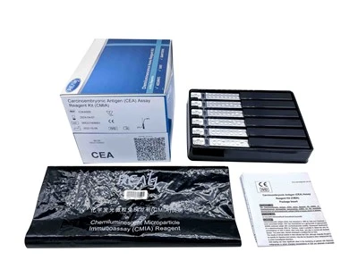
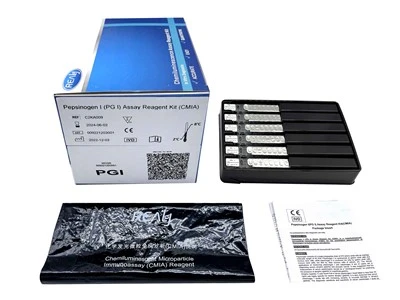
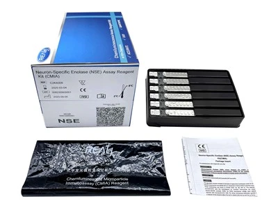
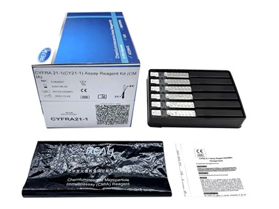
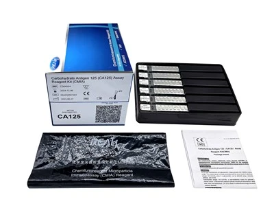
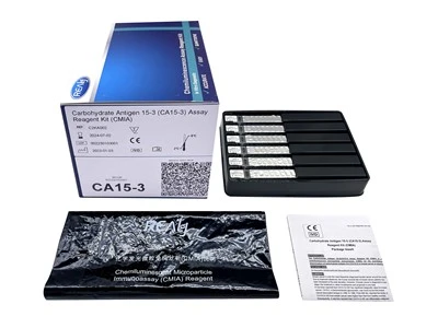
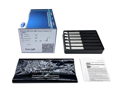
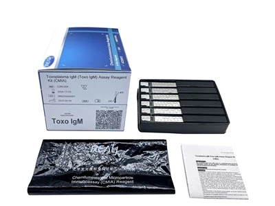
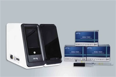
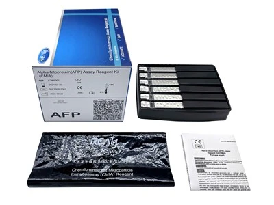
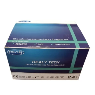
CLIA is a technique for determining sample concentrations based on the intensity of the light emitted by a chemical and biological reaction. The chemiluminescence (CL) systems and immunoreactions are combined in CLIA. Some chemicals have been used as CL labels, and the system generates chemiluminescence when the CL substrates are added, allowing the samples to be measured. The most frequent CL substrates include luminol, their derivatives, alkaline phosphatase (ALP), peroxidase, and acridinium ester compounds. In CLIA, the enzyme is also used for the markup of the target proteins. ALP and horseradish peroxidase are widely used for enzyme labeling.
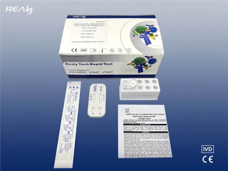
Chemiluminescence immunoassay contains two systems, immunoassay and chemiluminescence assay. The immunoassay system uses chemiluminescent substances or enzymes as markers, which are directly labeled on the antigen or antibody, and the antigen-antibody immune complex is formed through the reaction between the antigen and the antibody. The chemiluminescence analysis system is a luminescent substrate added with an oxidant or an enzyme after the immune reaction. After the chemiluminescent substance is oxidized by the oxidant, an intermediate in an excited state is formed, which emits photons to release energy to return to a stable ground state. The luminescence intensity can be detected using a luminescence signal measuring instrument. According to the relationship between the chemiluminescence marker and the luminescence intensity, the content of the analyte can be calculated using the standard curve.
Three Different Label Systems of CLIA
CLIA have three different label systems according to the difference of physical chemistry mechanism of the light emission:
Label Chemical Directly Involved in the Light Emission Reaction
This kind of chemical with special structure can transfer to an excited state through chemical reaction. Photons would be released when the chemical fell to ground state from the excited state. The typical chemical is acridinium ester and its derivatives. Exposure of an acridinium ester label to an alkaline hydrogen peroxide solution triggers a flash of light. A subsequent development has been the acridinium sulfonamide ester labels. It is also triggered by alkaline hydrogen peroxide to emit a flash of light.
Enzyme Catalyzed Light Emission Reaction
This type of chemiluminescence utilizes enzymes to label antibody. Technically speaking, it is an enzyme linked immunoassay that uses luminescent chemical as substrate instead of chromogen. The most widely used enzymes are horseradish peroxidase (HRP) and alkaline phosphatase (AP), each has its own luminescent substrates. Luminol is a very common chemiluminescent substrate used for detection of HRP. HRP catalyzes the decomposition of luminol in the presence of peroxide to produce an excited state intermediate. Flashes of visible light (maximum at 425nm) is emitted on decay of the singlet intermediate.
Redox Reaction Mediated Light Emission Reaction
Another CL system is noteworthy because the reagent is regenerated and thus can be recycled. This system utilizes ruthenium tris-bipyridine (bpy) as label, involves reaction of Ru(bpy)33+ and Ru(bpy)3+ to produce an excited state of Ru(bpy)32+, a stable species which decays to the ground state by emitting an 620 nm orange emission. Ru(bpy)33+ and Ru(bpy)3+ can be electrogenerated from Ru(bpy)32+ by reduction at approximately -1.3 V, and oxidation at approximately + 1.3 V. This system is dedicated for electrochemiluminescence with ultrahigh sensitivity and specificity.
Advantages of Chemiluminescence Immunoassay System
High Sensitivity
Sensitivity is the key to the superior performance of immunoassay analyzer. The sensitivity of some analyzers can reach 10 -16 mol / L (RIA for 10-12 mol / L). Another example is chemiluminescent substrates (such as AMPPD), the concentration of alkaline phosphatase can be detected than the chromogenic substrate to be sensitive 5 х 10 ^ 5 times.
Wide Linear Kinetic Range
The luminescence intensity of chemiluminescence immunoassay is linear between 4 and 6 orders of magnitude with respect to the concentration of the measured substance. This is a significant advantage compared to the absorbance (OD) range of 2.0 for colorimetric enzyme immunoassays.
Long Duration of Light Signal
The glow type CLIA produces a light signal that lasts for hours or even a day. This certainly simplifies the experimental operation and measurement.
The Analysis Method is Simple and Fast
Most of the chemiluminescence immunoassay analyzers are one-step mode that only requires the addition of one reagent (or a combination of reagents).
Stable Results, Small Error
The sample directly emits light on its own, without any light source irradiation, eliminating the impact of various possible factors (light source stability, light scattering, light wave selector, etc.) to the analysis. This makes the analysis results sensitive, stable and reliable.
Good Safety and Long Service Life
Chemiluminescent immunoassay analyzer eliminates the use of radioactive substances. So far, it has not been found to be harmful. Moreover, the reagents used for immunoassay are stable and can be stored for up to one year.
- 96-well microtiter plate and sealers
- Sample Diluent
- Capture and/or detection antibodies
- Avidin/HRP Conjugate
- Wash Buffer
- Substrate Solution
- Detailed Protocol
Applications of Chemiluminescence Immunoassay System
Clinical Diagnostics: CLIA is extensively used in clinical laboratories to diagnose various diseases and medical conditions. It can measure biomarkers, hormones, antibodies, and other analytes indicative of specific diseases.
Endocrinology: It is commonly used to assess thyroid function, reproductive hormone levels, and adrenal hormone levels, aiding in the diagnosis and management of endocrine disorders.
Therapeutic Drug Monitoring (TDM): CLIA is employed in TDM to measure drug concentrations in the blood, ensuring that medications are within the optimal therapeutic range.
Allergy and Immunology: CLIA is used to detect allergen-specific IgE antibodies in the blood, aiding in the diagnosis of allergies. It is also utilized to assess immune response and antibody levels in various immunological disorders.
Infectious Disease Testing: CLIA is widely used for detecting infectious agents, such as viruses, bacteria, and parasites, in clinical samples.
Autoimmune Disease Testing: CLIA is used to detect autoantibodies associated with autoimmune diseases, such as rheumatoid arthritis, systemic lupus erythematosus, and multiple sclerosis.
Oncology and Cancer Biomarkers: CLIA is applied in oncology to detect tumor markers and cancer-specific antigens.
Reproductive Health: CLIA is used in reproductive health to measure hormones related to fertility and pregnancy.
Drug Testing: CLIA is employed in drug testing programs, workplace drug screening, and forensic toxicology to detect drugs of abuse or therapeutic drugs in biological samples, such as urine and blood.
Medical Research: CLIA is an essential tool in medical research for quantifying biomolecules and studying disease mechanisms. It aids in understanding disease progression, identifying new biomarkers, and evaluating potential therapeutic interventions.
Procedure of Chemiluminescence Immunoassay System
Define the Expected Dynamic Range
It is important to figure out the approximate concentration range of the analyte in your sample. In addition, you need to figure out what the optimal concentration range is recommended in order to detect your desired analyte.
Define the Assayed Specimen
The type of specimen (e.g. blood, serum, urine, etc.) will help you understand the expected dynamic range of analyte contained within the specimen.
Select the Antibodies to be Used in the Assay
You will want to select antibodies that are highly specific for your unique analyte. Specificity is key to the success of your assay.
Choose Your Best Magnetic Beads
You will need to determine what the optimal bead size, bead surface area and iron oxide content needs to be for your specific analyte assay.
Optimize the Coating Procedure
When coating your beads, you will want to take into account the adsorption properties of the beads, any covalent binding you will need to create and the biological effects of linking proteins to your beads, or if you will need bio-activated ones.
Select Your Homogenization Method
Your choice of homogenization will depend on the equipment you have, the personnel who work on the equipment, the size of your samples and the ease of use within the assay.
Select the Correct Biomagnetic Separation Technology
This technology helps removing free antibodies and other contaminants. Systems that can help you easily validate your process and can help you scale up your process are preferred.
Enhance Your Immunoassays When Required
If you can make your assay better, it is important to do so. Making your assay more efficient, more accurate and easier to perform is important to the final results of your analysis.
1.The default concentration unit of this assay is ng/mL or nmol/L.
Conversion factor:
- o ng/mL x 2.5=nmol/L
- o nmol/L x 0.4= ng/mL
2.Due to methodological differences or antibody specificity, there may be deviations between the test results of reagents from different manufacturers. Therefore, direct comparisons should not be made to avoid false interpretation.
3.When the 25-OH VD concentration in the sample exceeds 100.00 ng/mL, a sample dilution could be performed before measurement (recommended 2-times dilution).
4.Any result below the minimum detection limit will be reported as <3.00 ng/mL; any result above the maximum detection limit will be reported as >100.00 ng/mL.
Handling Precautions
Do not use reagent kits beyond the expiration date.
Do not interchange reagent components from different reagents or lots.
Prior to loading the Reagent Kit on the system for the first time, the Reagent Kit requires mixing to re-suspend magnetic microbeads that have settled during shipment. For magnetic microbeads mixing instructions, refer to the Preparation of the Reagent section of this package insert.
To avoid contamination, wear clean gloves when operating with a reagent kit and sample.
Over time, residual liquids may dry on the septum surface. These are typically dried salts which have no effect on assay efficacy.
To avoid evaporation of the liquid in the opened reagent kits in a refrigerator, it is recommended that the opened reagent kits be sealed with reagent seals contained within the packaging. The reagent seals are "single use," and if more seals are needed, please contact us.
Differences Between ELISA and CLIA Assays
We can characterize the differences between these types of assays based on the type of substrate and signal they use, how sensible they are, or even the advantages and disadvantages of each test.
Type of substrate and signal
The substrate of an immunoassay is a key piece of the reaction, being the one that allows us to measure the results with ease and precision.
While an ELISA test utilizes chromogenic substrates like TMB or ABTS, which look to tint the sample to a greater or lesser degree to be able to measure the colour change; the CLIA assays utilize luminescent substrates like Luminol or acridinium ester, that glow after the reaction allowing to measure the light given by it.
Differences on sensibility
The difference between the grade of sensibility on both types of assays is stark. Despite both being able to detect fairly small quantities of the desired protein and are highly precise by their own merit, in this particular regard, CLIA tests are the clear winners.
Where an ELISA can detect proteins in a concentration of down to 500pg/mL, a CLIA test can detect them even in a concentration of 1pg/mL, making this technique an ultrasensitive one.
Advantages and disadvantages
Each one of these assays has a multitude of advantages and disadvantages that make them perfect for different kinds of experiments.
ELISA tests, for example, are fast and easy to perform in their most standard form, and many brands have on sale specialized kits that reduce the time needed to perform the protocol even further. They are also widely used in labs all over the world, making them a standard assay and a must-learn for every budding researcher. Running against them, ELISA kits have a fixed period of time for the reading to happen, as it has to be done in the moments following the reaction enzyme-substrate, making it difficult to use them along flexible planning.
On the other hand, CLIA assays have a wider dynamic range than their counterparts, so they are considerably more precise. Their highly stable reagents and low depth ratio make them easy to automate and allow making the pertinent readings in a wide period of time after completing the assay. However, CLIA assays make use of highly specialized equipment, like a luminescence microplate reader, making it a costly endeavour that is not widely accessible by the average laboratory.
Designing CLIA assays requires the consideration of different aspects, encompassing the raw materials for the reagent development & methods selection, together with the choice of the assay format. Material suppliers are a key factor for a successful design and development of an assay. An ideal supplier should be able to provide required raw materials not only at a reliable cost but also available to provide the required bulk quantities for scaling up the reagent. Moreover, suppliers should provide different lots to assess the lot to lot variability to check the impact in the assay to be developed.
Strategies for Choosing Magnetic Beads
Magnetic beads are available in different sizes, ranging from nanometers to a few micrometers. Smaller beads will likely require more time to separate using a magnetic separator. The separation time of the magnetic beads depends strongly on the amount of the magnetic pigment. Higher pigment percentage will result in faster magnetic bead separation. A balance between the magnetic pigment and the size of the magnetic beads should be reached to maintain the correct density of the magnetic beads.
● Chemical Modified Magnetic Beads for CLIA
A simple but less stable strategy for conjugating a magnetic bead surface can be achieved through passive adsorption of the antigen or antibody directly on the surface of a “plain” magnetic bead. Alternatively, the modification of the surface chemistry of the magnetic beads is a great option if covalent attachment of the antigen or antibody is important for the assay. Active groups take advantage of the large surface area of the magnetic beads and provide abundant anchoring points for your antibodies or antigens to become covalently conjugated to the bead. Some examples of pre-activated chemicals that can be used are the popular Tosyl group or epoxy. There are other groups that are used which require activation, such as carboxyl (-COOH) groups, amines (-NH2), and hydroxyls (-OH). This practice is also known as the chemical functionalization of magnetic beads.
● Biological Modified Magnetic Beads for CLIA
Biological modified magnetic beads (also known as surface biofunctionalization) are also a common strategy for CLIA development. There are several types of biological modified magnetic beads. The antibody or antigen of choice can be modified with a biotin linker which binds to a magnetic bead modified with streptavidin. Protein A binds the Fc region of most immunoglobulins making it an attractive choice for the immobilization of antibodies to the magnetic beads. Similarly, Protein G binds either the Fc or Fab region of a spectrum of immunoglobulins and can be used to immobilize antibodies. Sometimes Protein A and G are used together to increase the spectrum of potential antibody binding. It is also possible to conjugate specific proteins but this requires optimization.
When biological modified magnetic beads are selected for a CLIA assay it is important to check the compatibility of these beads with the sample type, in order to prevent non-specific interactions with components of the sample with the biological surface of the magnetic beads.
Our certificate
We have passed various certificates such as CE and ISO, and the products we provide have quality assurance.


Common Problems of Chemiluminescence Immunoassay System
As one of the most professional chemiluminescence immunoassay system manufacturers and suppliers in China, we're featured by quality products and good service. Please rest assured to wholesale cheap chemiluminescence immunoassay system from our factory. Contact us for more details.
chemiluminescence immunoassay for biomarker translocation, chemiluminescence immunoassay for biomarker precipitation, chemiluminescence immunoassay for drug integration